
BLOG
The Ultimate Clinic Guide to Photographic Packing Evidence: Efficacy, Wavelengths, and ROI
2026年9月15日
Why Photographic Packing Evidence Is the Missing Final Step in Aesthetic Quality Control
In the competitive world of medical aesthetics, clinics invest heavily in FDA-cleared and Medical CE certified laser platforms, train staff on selective photothermolysis, and meticulously calibrate parameters such as fluence, pulse width, and spot size. Yet a critical gap remains: the lack of standardized photographic packing evidence as a final quality control checkpoint. This article defines the protocol, technical requirements, and business impact of implementing photographic packing evidence as the last step before patient discharge.
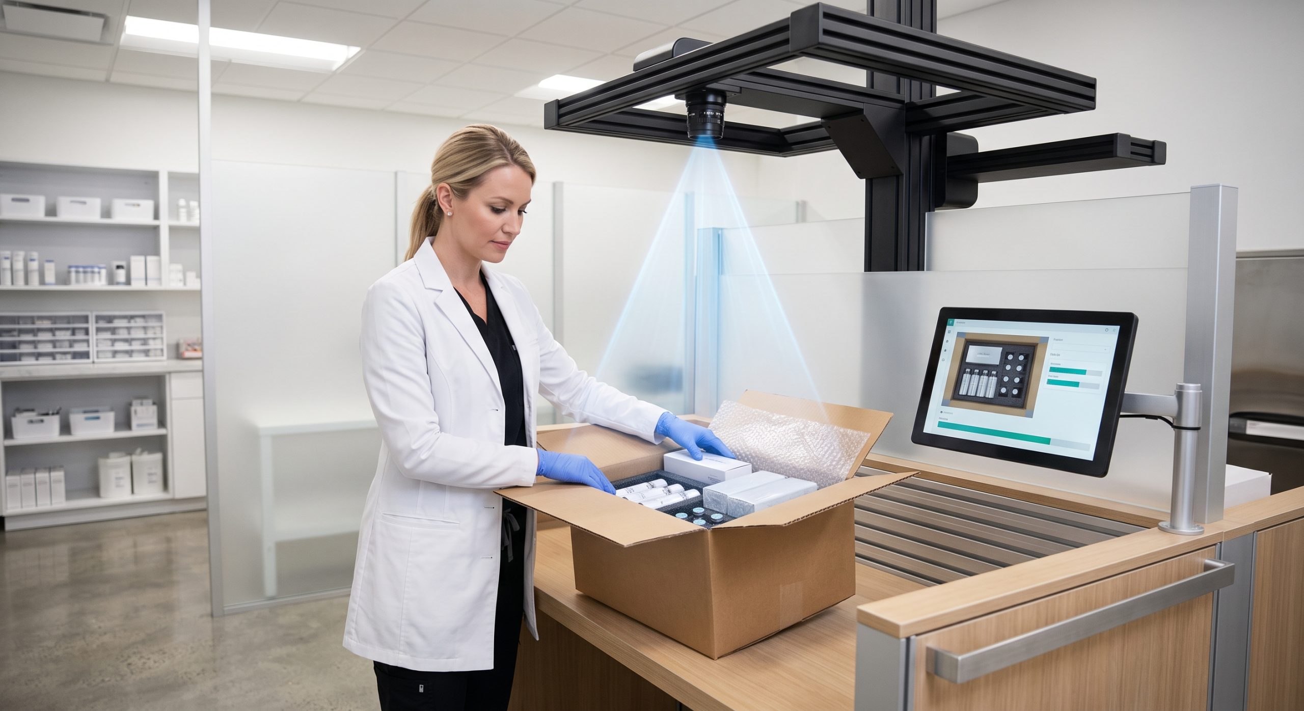
What Is Photographic Packing Evidence?
Photographic packing evidence refers to the standardized, metadata-rich image set captured immediately after a laser or energy-based treatment, before the patient leaves the clinic. It is not a marketing before/after photo. It is a clinical quality control artifact that documents:
- Immediate tissue response (e.g., perifollicular edema, erythema, or graying of hair).
- Device settings used: wavelength (755nm, 808nm, 1064nm), fluence (J/cm²), pulse width (ms), spot size (mm), and cooling temperature.
- Anatomical site, Fitzpatrick skin type, and treatment grid pattern.
- Timestamp, operator ID, and device serial number.
This evidence pack becomes the legal and clinical baseline for any adverse event investigation, insurance claim, or outcome dispute.
Technical Foundation: Why Wavelength and Cooling Dictate Evidence Quality
To understand what constitutes valid photographic packing evidence, one must first understand the physics of the device. Modern diode laser platforms for hair removal and vascular/pigment treatment typically combine 755nm (melanin affinity), 808nm (balanced depth and melanin absorption), and 1064nm (deep penetration, safer for darker skin). The spot size directly affects penetration depth and scattering. A larger spot size (e.g., 10mm–12mm) with high fluence (e.g., 10–40 J/cm²) and short pulse width (e.g., 10–100 ms) produces more reliable thermal damage to the follicle.
However, without sapphire contact cooling or TEC cooling at 0–5°C, epidermal injury risk rises, making photographic evidence of skin reaction essential. The evidence must capture the epidermal preservation alongside the target response.
| Key Parameter | Technical Specification |
|---|---|
| Wavelength / Laser Type | 755nm / 808nm / 1064nm Diode |
| Spot Size | 10mm – 12mm (adjustable) |
| Energy Density / Fluence | 10 – 40 J/cm² |
| Pulse Width | 10 – 100 ms |
| Cooling System | Sapphire Contact Cooling (0–5°C) / TEC |
| Certification | Medical CE, FDA Cleared, ISO 13485 |
Implementing the Evidence Pack: A Step-by-Step Protocol
Step 1: Standardized Capture Setup
- Use a cross-polarized or parallel-polarized photographic system to eliminate surface glare and reveal vascular or pigmentary changes.
- Fixed focal length, distance, and lighting (e.g., 5500K color temperature).
- Include a calibration target (e.g., color checker and ruler) in every frame.
Step 2: Metadata Embedding
Each image must be tagged with DICOM-like metadata: device model, laser bar origin (e.g., imported German or US bars), handpiece serial number, shot count, and coolant temperature. This aligns with ISO 13485 traceability requirements.
Step 3: Immediate Clinical Review
Before the patient leaves, the operator reviews the evidence for expected endpoints. Absence of expected perifollicular edema may indicate insufficient fluence or poor contact cooling. Presence of blanching or vesiculation requires immediate intervention and documentation.
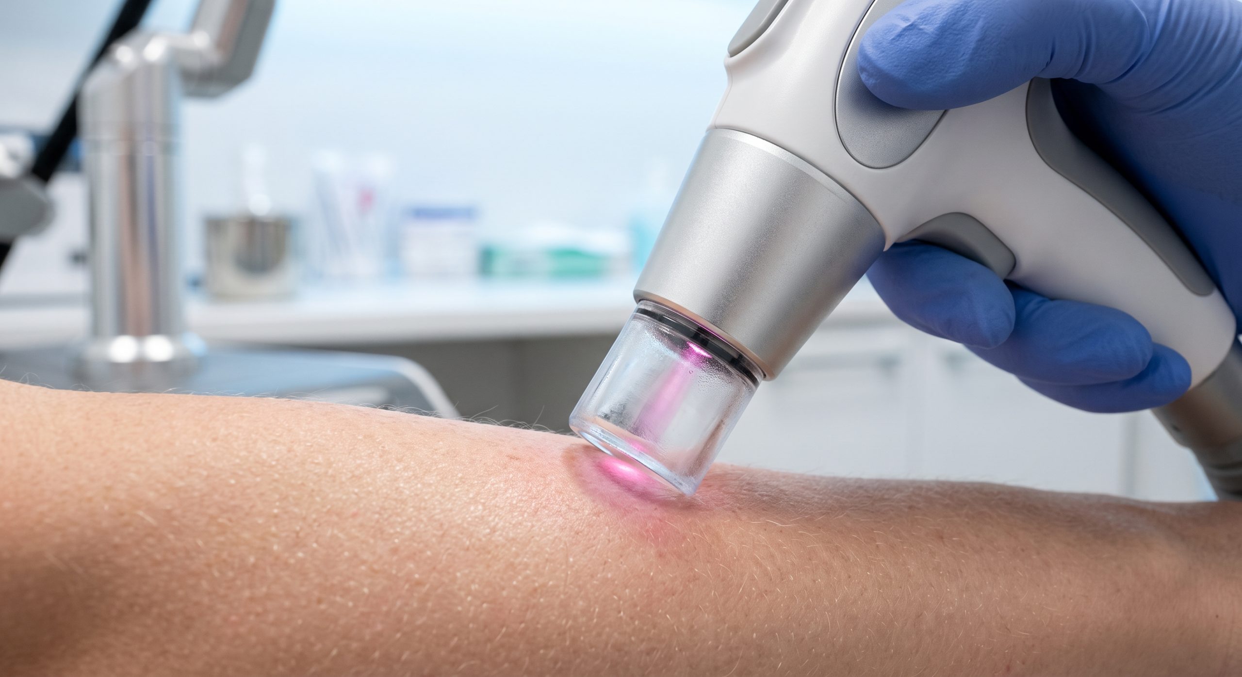
Fitzpatrick-Specific Evidence Interpretation
For Fitzpatrick I–III, expected evidence includes mild erythema and perifollicular edema. For Fitzpatrick IV–VI, the evidence must show minimal epidermal change and no post-inflammatory hyperpigmentation (PIH). The photographic pack must be compared against a baseline taken pre-treatment under identical conditions. Clinics using 755nm/808nm/1064nm platforms can adjust fluence and pulse width based on this evidence, creating a feedback loop for continuous protocol optimization.
Business & Compliance Impact: ROI of Photographic Packing Evidence
- Risk mitigation: Reduces malpractice claims by providing objective proof of appropriate technique and settings.
- Insurance & Medical CE audits: Demonstrates compliance with ISO 13485 and FDA post-market surveillance expectations.
- Patient retention: Sharing controlled evidence builds trust and justifies premium pricing.
- Operational efficiency: Identifies underperforming handpieces or degraded laser bars before patient complaints arise.
For a clinic performing 20 treatments per day, a 5-minute evidence pack adds 100 minutes of labor daily but can reduce complication-related revenue loss by an estimated 15–30% annually.
Conclusion: The Last Step That Protects Everything Before It
Photographic packing evidence is not an administrative burden; it is the final quality control seal on a chain that begins with device certification (Medical CE, FDA, ISO 13485) and continues through wavelength selection, fluence optimization, and cooling system performance. Clinics that adopt this step as standard protocol will outperform peers in safety, compliance, and patient satisfaction. The evidence pack is the difference between assuming quality and proving it.